Features

Our Services

Magnetic Resonance (MRI)

MRI stands for Magnetic Resonance Imaging,a non-invasive diagnostic procedure that uses magnetic fields and radio frequencies to generate detailed anatomical and functional images. MRI scans have an advantage over some forms of scanning because they can image different types of organ tissue without ionizing radiation. More traditional forms of imaging, such as X-rays, are limited in how much tissue they can image.

Computerized Tomo (CT)

A CT or CAT scan, also known as a computed tomography scan, is a medical imaging method that uses special X-ray equipment to obtain cross-sectional pictures of the body. The CT computer displays these pictures as detailed images of organs, bones, and other tissues. This type of imaging can be very helpful in diagnosing injuries or fractures or in identifying disease in its earliest stages.

Roentgen (X-Ray)

An x-ray (radiograph) is a noninvasive medical test that helps physicians diagnose and treat medical conditions. X-Ray images are used to detect fractured bones, signs of arthritis and other potential internal complications. The rays pass through the body at different rates depending upon the density of the tissue. Soft tissues such as muscle and fat are passed through much easier than harder matter such as bone for example. The rays that pass through the body are captured by the x-ray machine to provide an image of the body’s internal structure.

Ultrasound (US)

Ultrasound imaging (sonography) uses high-frequency sound waves to view inside the body. Because ultrasound images are captured in real-time, they can also show movement of the body's internal organs as well as blood flowing through the blood vessels. Ultrasound imaging is a noninvasive medical test that helps physicians diagnose and treat medical conditions.

Electroencephalogram (EEG)

An electroencephalogram (EEG) is a test used to find problems related to electrical activity of the brain. An EEG tracks and records brain wave patterns. Small metal discs with thin wires (electrodes) are placed on the scalp, and then send signals to a computer to record the results.

Electromyoneurography(EMNG)

Electromyoneurography (EMNG) is a diagnostic procedure to assess the health of muscles and the nerve cells that control them (motor neurons). Motor neurons transmit electrical signals that cause muscles to contract. ... An EMG uses tiny devices called electrodes to transmit or detect electrical signals.

Why Choose Us

img
img
img
img
img
img

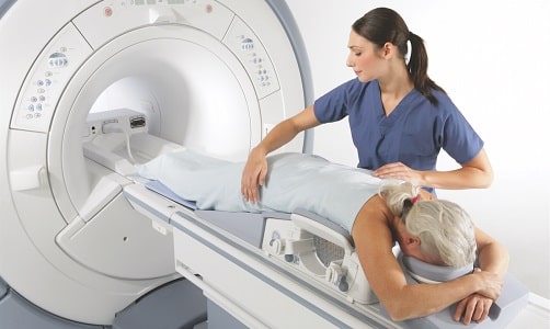
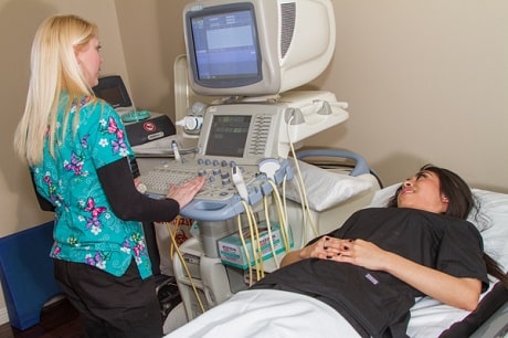
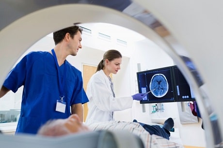
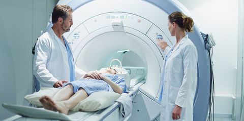
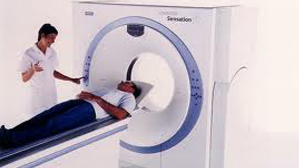
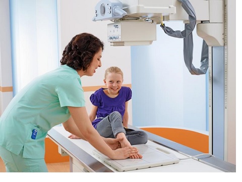
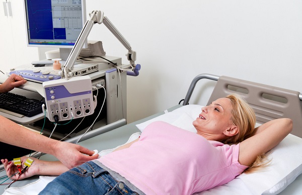
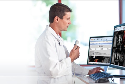
Great Infrastructure

State of the art equipments under a well structured enviroment produces best results.
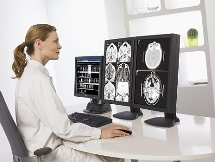
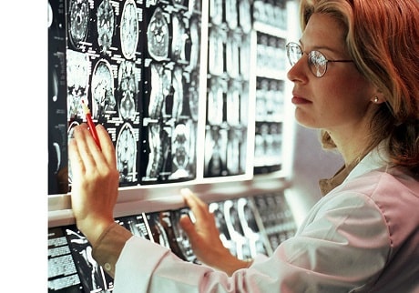
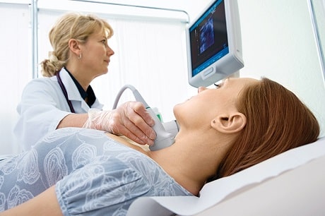
Qualified Doctors

Experienced local radiologists that are well known in their areas and professors from abroad for consultation.

Quality Service

In IHC, our priority is quality. No matter it is a simple procedure or a complex one, we take it seriously.

4500
Exams per year
86
Imaging Procedures
21
Advanced Techniques
3000
Happy Patients

Our Departments

img

Our MRI department is equipped with a GE Signa HD, 1.5T MRI scanner.

The Signa HDe 1.5T High-field MRI is engineered for enhanced image contrast, reduced blurring and reduced artifacts so your doctor can see more. This system also incorporates motion correction to help reduce the need for rescans. GE Signa HDe is a high resolution, whole body imaging system known for its excellent performance. The powerful magnetic resonance imaging platform provides high definition results and a comprehensive range of applications.

img

Computed Tomography (CT) is a diagnostic procedure that uses special X-ray equipment to obtain cross-sectional pictures of the body. The CT computer displays these pictures as detailed images of organs, bones and other tissues. Radiation exposure is minimized with our Siemens 64 Slice multi-detector Sensation CT Scanner at IHC. The multi-slice technology reduces radiation significantly. The scanner, in real time, alters the radiation emitted, depending on the thickness of the body part being scanned.

img

With our state of art X-Ray imager, and CR unit, we are able to do varous X-Ray exams at our X-Ray department. Fully digitized, DICOM images, ready for reporting and review in matter of seconds, and available for the patient and referral doctor.

img

With real-time ultrasound images, we are able to see movement of internal tissues and organs, and enable physicians to see blood flow and heart valve functions. This helps to diagnose a variety of heart conditions and to assess damage after a heart attack or other illness. With the state of art ultrasound scanner with various multi-purpose probes, ultrasound exams are fast, painless, and stress-free in our Ultrasound department.

img

Well known doctors working with high-end equipment, our EEG/EMG department produce accurate test result for our patients, resulting satisfied happy patients.

How we work

Appointment

Most of the MRI, CT, Ultrasound and X-Ray exams require special conditions to be met by the patient and secific preperations of the equipment. For this reason, it is necessary to plan the daily schedule with patient appointments. This prevents unnecessary waiting periods for the patients and more effective use of the equipment.

img

Inform the Patient

Before every exam, we inform our patients about the details of the exam. What will be the procedure we will follow, how will the patient feel during the exam, necessity of any extra medicaitons, etc etc. Patients take their exams with the full understanding about it.

img

Exam

Medical Imaging procedures are complex and need full dedication to be able to obtain the best images. Our experienced and knowledgable operators and doctors perform every procedure with extra care, so that the results are crystal clear.

img

Remote Consultation

It is always good to have a second opinion! No matter how experienced our local doctors are, we always prefer to take the opinion of professors from Turkey, who are known world wide in their specific areas of expertise. This ensures the accuracy of our reports.

img

Final Patient Report

We are always proud of the quality of our final patient reports. With the help of high tech equipment, our doctors are able to see more details in images. On top of this, sharing the patient images with our consultant professors abroad, no room is left for error. Our accurate reports help physicians to decide correct diagnosis.

img