General Radiology
Radiography, or an x-ray, as it is most commonly known, is the oldest and most frequently used form of medical imaging. Discovered more than a century ago, x-rays can produce diagnostic images of the human body on film or digitally on a computer screen.
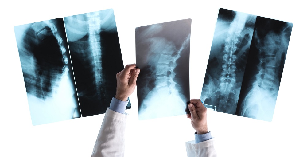
X-ray imaging is the fastest and easiest way for a physician to view and assess broken bones, such as skull fractures and spine injuries. At least two images (from different angles) are taken and often three images are needed if the problem is around a joint (knee, elbow or wrist). X-rays also play a key role in guiding orthopedic surgery and in the treatment of sports-related injuries. X-ray may uncover more advanced forms of cancer in bones, although early screening for cancer findings requires other methods.
What is the purpose of an X-Ray?
X-Ray images are used to detect fractured bones, signs of arthritis and other potential internal complications. The rays pass through the body at different rates depending upon the density of the tissue. Soft tissues such as muscle and fat are passed through much easier than harder matter such as bone for example. The rays that pass through the body are captured by the x-ray machine to provide an image of the body’s internal structure.
Your physician may order an X-ray to learn more about:
- Bones, including fractures, arthritis and bone cancer
- Lung infections and lung cancer
- Problems in your digestive tract
- Blood vessel health
- Signs of congestive heart failure, such as an enlarged heart
Do I need any preparation for this examination?
Before you have your x-ray you may be required to remove parts of your clothing and wear a gown to ensure accurate images. The part of your body that is being observed is then placed in front of a cassette plate whilst the Radiographer lines up the x-ray machine. They will then stand behind a protective glass screen whilst your image is taken.
What happens after the examination?
Once the Radiographer is satisfied that the images are accurate the procedure is finished. The taking of the images takes only a few seconds whilst the construction of the films may take several minutes. There is no pain or discomfort caused by the x-rays and you will have your films complete with Radiologist’s report within half an hour from walking into the building in most cases. Remember to take you film along with your report to your next doctor’s appointment.
Chest X-Ray
A chest x-ray is typically performed as the first imaging test for symptoms of shortness of breath, a bad or persistent cough, chest pain, chest injury or fever. Individuals with known or suspected medical conditions such as congestive heart failure or cancer may undergo chest x-rays to follow their response to treatment, or to determine changes that would require a change in their medical management.
X-Ray Services offered
- Chest
- Spine
- Skull
- Sinus
- Ribs
- Pelvis
- Extremity
- etc.
If you are pregnant, please notify your physician.