Electromyography (EMG)
Electromyography (EMG) is a diagnostic procedure that evaluates the health condition of muscles and the nerve cells that control them. These nerve cells are known as motor neurons. They transmit electrical signals that cause muscles to contract and relax. An EMG translates these signals into graphs or numbers, helping doctors to make a diagnosis.
A doctor will usually order an EMG when someone is showing symptoms of a muscle or nerve disorder. These symptoms may include tingling, numbness, or unexplained weakness in the limbs. EMG results can help the doctor diagnose muscle disorders, nerve disorders, and disorders affecting the connection between nerves and muscles.
What is the purpose of an EMG Test?
Your doctor may perform an EMG if you’re experiencing symptoms that may indicate a muscle or nerve disorder. Some symptoms that may call for an EMG include:
- tingling
- numbness
- muscle weakness
- muscle pain or cramping
- paralysis
- involuntary muscle twitching (or tics)
The results of an EMG can help your doctor determine the underlying cause of these symptoms.
How Do I Prepare for an EMG?
Make sure to notify your doctor about any over-the-counter or prescription medications you may be taking. It’s also important to tell your doctor if you have a bleeding disorder, or if you have a pacemaker or implantable defibrillator. You may not be able to have an EMG if you have any of these medical conditions or devices.
If you are able to have an EMG, you should do the following beforehand:
- Avoid smoking for at least three hours before the procedure.
- Bathe or take a shower to remove any oils from the skin. Don’t apply any lotions or creams after washing.
- Wear comfortable clothing that doesn’t obstruct the area that your doctor will be evaluating. You may be asked to change into a hospital gown right before the procedure.
What should I expect during my EMG Test?
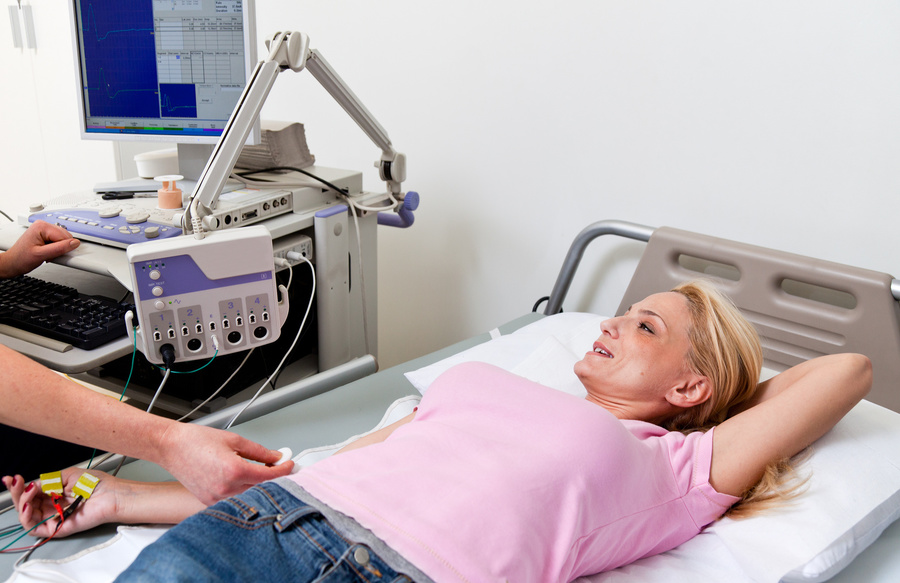
You will be asked to lie down on an examination table or to sit in a reclined chair. Your doctor may ask you to move into different positions during the procedure.
An EMG has two parts: the nerve conduction study and the needle EMG. The nerve conduction study is performed first. During this portion of the procedure, your doctor will apply several electrodes to the surface of your skin, usually in the area where you are experiencing symptoms. These electrodes will evaluate how well your motor neurons communicate with your muscles. Once the test is complete, the electrodes are removed from the skin.
After the nerve conduction study, your doctor will perform the needle EMG. Your doctor will first clean the affected area with an antiseptic. Then, they will use a needle to insert electrodes into your muscle tissue. You may feel slight discomfort or pain while the needle is being inserted.
The needle electrodes will evaluate the electrical activity of your muscles when contracted and when at rest. These electrodes will be removed after the test is over.
During both parts of the EMG procedure, the electrodes will deliver tiny electrical signals to your nerves. A computer will translate these signals into graphs or numerical values that can be interpreted by your doctor. The entire procedure should take between 30 and 60 minutes.
What do my EMG test results mean?
Your doctor may review the results with you right after the procedure. However, if another health care provider ordered the EMG, then you may not know the results until you attend a follow-up appointment with your doctor.
If your EMG shows any electrical activity in a resting muscle, then you may have:
- a muscle disorder
- a disorder affecting the nerves that connect to the muscle
- inflammation caused by an injury
If your EMG shows abnormal electrical activity when a muscle contracts, then you may have a herniated disc or a nerve disorder, such as ALS or carpal tunnel syndrome.
Depending on your results, your doctor will talk to you about any additional tests or treatments that might be needed.