- January
15
2021 - 150
- 0
MRI and its use in several medical conditions
The development of the MRI scan represents a huge milestone for the medical world.
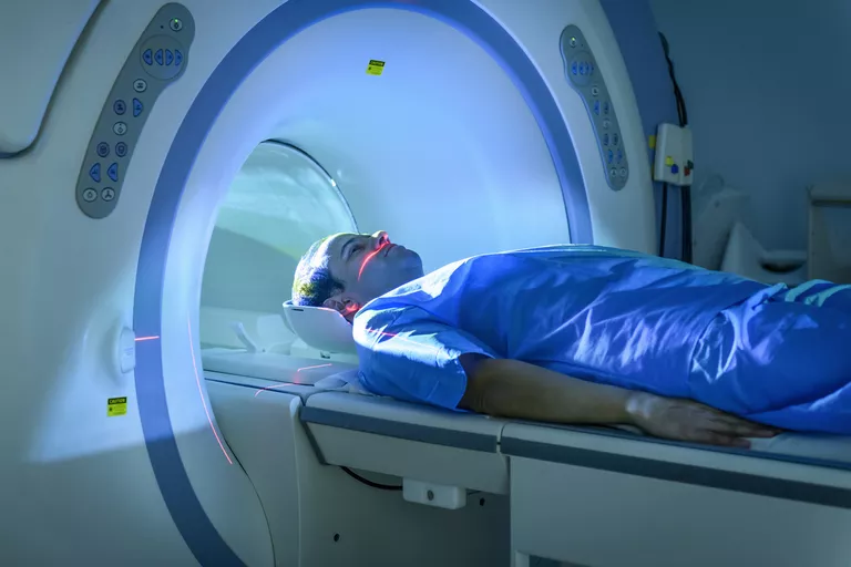
MRI technology was developed because of the need for brain imaging. It is one of the few imaging tools that can see through bone (the skull) and deliver high quality pictures of the brain's delicate soft tissue structures. Its greatest innovation is allowing us to see which parts of the brain have been damaged while a patient is still alive. In the past, we had to wait until a post-mortem to find out. It helps physicians evaluate both the structure of the brain, its normal functional anatomy and how it is working.
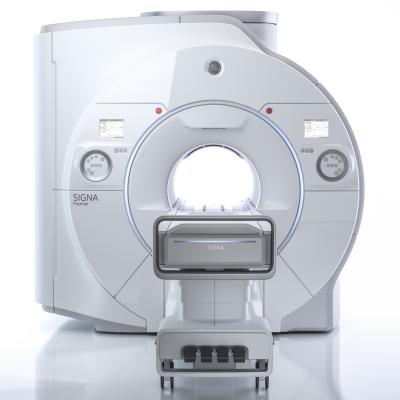
Fig 1. Magnetic Resonance Scanner
MRI (Magnetic Resonance Imaging) is a non-invasive imaging technique that does not involve exposure to ionizing radiation and isn't painful.
It uses a magnetic field to measure the amount of water in the tissues. The human body is largely made of water molecules, which are comprised of hydrogen nuclei (protons), and oxygen atoms, they become aligned in a magnetic field and create a signal that is processed to form an image of the body.

Figure 2. Water molecules and their importance for MRI
Medical conditions where it is extremely helpful:
Acute ischemic infarct - Ischemic stroke is the most common neurological cause of severe disability and death. MRI is considered more useful than a CT scan for diagnosing acute ischemic stroke within 12 hours of a person's first stroke symptom and then be able to decide the treatment plan.
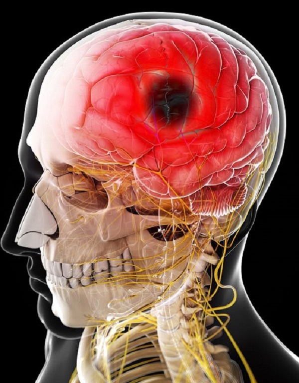
Figure 3. Depiciting an ichemic stroke of the brain
Head trauma - MRI is very useful in the detection of all types of intracranial haemorrhage. It can detect signs of injury such as minute bleeding (microhemorrhage), small areas of bruising (contusion) or scarring (gliosis), which are invisible to the CT scan.
Multiple Sclerosis - MRI is recognised as the preferred and most sensitive imaging technique for the diagnosis of multiple sclerosis (MS). It has a proven capacity to define the plaques of multiple sclerosis provided by no other technique.
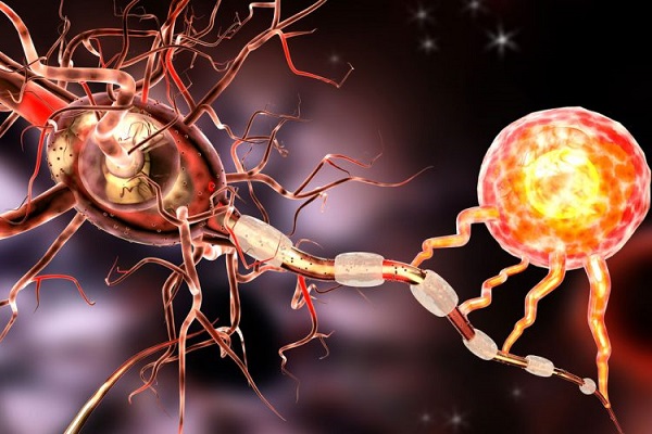
Figure 4. Depicting the neuronal cells and the immune system cells attacking their myelin sheaths in Multiple Sclerosis
Knee conditions - Bone fractures, arthritis, infections, effusions, trauma-related injuries. Due to the excellent contrast resolution of soft tissues, MRI demonstrates muscle and ligament tears and hematomas well.
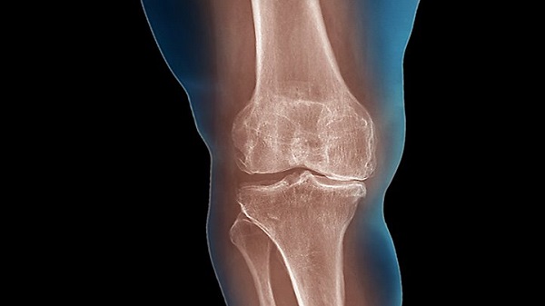
Figure 5. Knee joint articulation
Pancreas and biliary tract diseases - MRCP has virtually replaced ERCP as the primary investigative modality in all cases of obstructive jaundice not requiring early endoscopic intervention. Magnetic resonance (MR) imaging besides being non-invasive has the advantages of allowing detailed evaluation of the pancreatico-biliary tract with a large field of view and excellent patient tolerance.
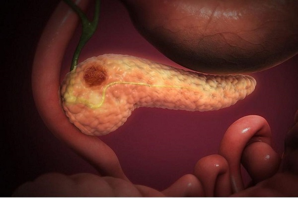
Figure 6 . Depicting the pancreas and its damaged parenchyma
Vascular malformations - Vascular malformations are benign (non-cancerous) lesions, often present at birth and composed of blood vessels that can cause functional or cosmetic problems. MRI is exquisitely sensitive to blood flow circulation and has proven particularly effective in the detection and localisation of vascular malformations.
Tumors of the spinal cord - MRI is capable of demonstrating the entire spinal cord and of differentiating solid from cystic intramedullary tumours and their local extent can be staged best by MRI.
Herniated discs - MRI is particularly useful for identifying and evaluating degenerated or herniated spinal discs. It can also be used to determine the condition of nerve tissue within the spinal cord.
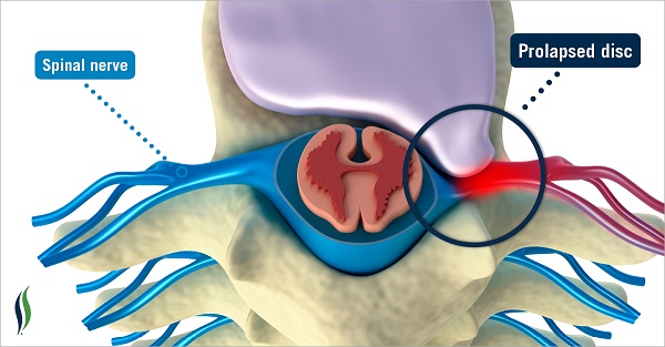
Figure 7. Depicting a prolapsed disc
Carotid Artery MRI - The carotid arteries are the two major arteries on either side of the neck that carry blood into the head and brain. Plaque buildup in the carotid arteries often mirrors that in the coronary arteries, but the carotids are more easily imaged, making them potentially useful vessels for the assessment of the risk of strokes, heart attacks and other adverse cardiovascular events. Carotid artery plaque revealed on MRI may be better indicator of cardiovascular disease than ultrasound-based carotid intima-media thickness test.
The carotid artery serves as window into the cardiovascular system.
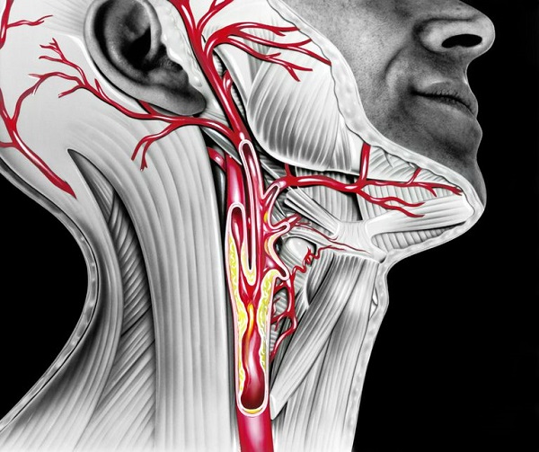
Figure 8. Depicting the carotid arteries and the plaque build up
High-resolution MRI can tell us the stage of plaque in the wall and tell us about plaque features that could lead to stroke.
Atherosclerosis - Atherosclerotic disease is characterized by a narrowing of the arteries caused by plaque buildup. Magnetic resonance angiography offers radiation-free method to detect atherosclerosis throughout the body.
With whole-body MRA, we are able to detect early atherosclerotic disease throughout the body, disease that would have been missed by methods that evaluate only a single vascular territory.
Thrombosis of the extremities - Deep vein thrombosis (DVT) occurs when a blood clot (thrombus) forms in one or more of the deep veins in your body, usually in your legs. Deep vein thrombosis can be very serious because blood clots in your veins can break loose, travel through your bloodstream and lodge in your lungs, blocking blood flow (pulmonary embolism). Magnetic Resonance Angiography (MRA) is a non-contrast-enhanced procedure that can visualize a thrombus directly by the visualization of methemoglobin, which is formed in a fresh blood clotso it has been proven to be accurate in diagnosing deep venous thrombosis (DVT) of the leg.
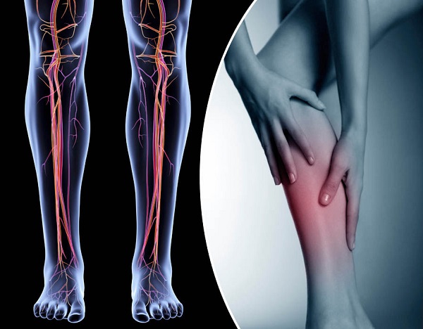
Figure 9.
MRI examination has no known long-term harmful effects, provided the safety precautions are followed. MRI does not use ionising radiation. Avoiding the need for exposure to ionising radiation (X-rays) is of significant benefit to younger people and children, and MRI can also be used safely in pregnancy, if required.
The benefits of this medical imaging are extensive, and include:
- Rapid and accurate diagnosis
- More evidence-based decision-making
- More personalized and better treatment
- Increased understanding of the effect of treatments on diseases
- Less recurrence of disease
For the patient, these benefits translate into a higher quality of life, the potential to enjoy a longer life span, and much less time to get back into a regular routine than would be required otherwise.